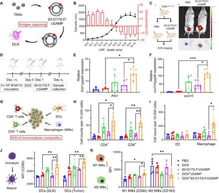
Fig. 5. LNP enhances STING activation and shifts immunocellular composition of the tumor microenvironment in vivo.

(A) Capture of the tumor antigens by 93-O17S-F. (B) The diameters and zeta potentials of 93-O17S-F and tumor lysate complex at different weight ratio. (C) Enhanced delivery of OVAAlexa647 to DLNs after being captured by 93-O17S-F in vivo. Photo credit: J.J. Chen, Tufts University. (D) The route of the in vivo STING activation experiments. (E and F) The relative expression of ifnb1 and cxcl10 genes in B16F10 tumors after the administration of 93-O17S-F/cGAMP for 6 hours. n = 6, *P ≤ 0.05 and ***P ≤ 0.001. (G) The activation of STING pathway recruited the immune cells to tumor sites. (H) The cell numbers of CD4+ and CD8+ T cells at tumor sites after the administration of 93-O17S-F/cGAMP for 48 hours. n = 5, *P ≤ 0.05 and **P ≤ 0.01. (I) The cell numbers of dendritic cells (DCs) and macrophages at tumor sites after the administration of 93-O17S-F/cGAMP for 48 hours. n = 5, *P ≤ 0.05. (J) The MFI of CD80 expressed on CD11c+MHC II+ DCs at DLNs and tumor sites. n = 5, **P ≤ 0.01. (K) The polarization of macrophages at tumor site determined by the MFI of CD80 and CD163 among CD11b+F4/80+ cells. n = 5, *P ≤ 0.05 and **P ≤ 0.01.
