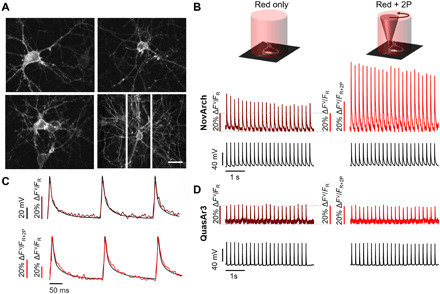
Fig. 4. 2P-activated voltage imaging in cultured rat hippocampal neurons.

(A) Fluorescence of Citrine in NovArch-Citrine showed good membrane trafficking. Scale bar, 50 μm. (B) Simultaneous voltage and fluorescence recordings of action potentials evoked through current clamp stimulation via patch pipette. Application of 2P illumination scanned around the cell periphery enhanced the NovArch fluorescence. Vertical scale bars show fluorescence normalized by either red-only illumination (FR) or red +2P illumination (FR+2P). The increased amplitude of the spikes under 2P illumination reflects an increase in overall brightness of the reporter (see, e.g., Fig. 3E). (C) The fluorescence recordings show close correspondence to simultaneous manual patch-clamp recordings with (top) red-only illumination or (bottom) red + 2P illumination. (D) QuasAr3 also resolved action potentials but did not show 2P photoactivation. Data are representative recordings from n = 6 neurons expressing NovArch and n = 6 neurons expressing QuasAr3.
