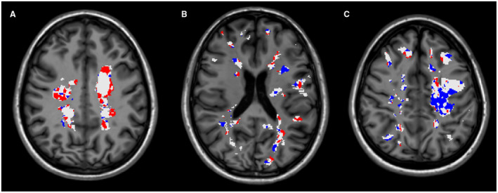
Figure 1.

Three single‐patient maps of lesional myelin content changes showing demyelinating (in red) and remyelinating (in blue) voxels derived from longitudinal [11C]PIB PET, localized inside white matter (WM) lesions (in white), overlaid onto the corresponding MPRAGE scans. A. Map of lesional myelin content changes of a 20‐year‐old woman with a disease duration of 3 years, where a clear prevalence of demyelination over remyelination is visible. B. The map of myelin content changes of this 27‐year‐old man with a 10‐year history of MS shows active demyelination together with moderate remyelination in all visible WM lesions. C. An extensive process of remyelination characterizes the map of myelin content changes of this 32‐year‐old woman with a disease duration of 3 years. Of note, the extensive lesion visible in the left‐hemispheric WM corresponds to a recent lesional area, characterized by large gadolinium‐enhancing regions on the corresponding T1 spin‐echo scans.
