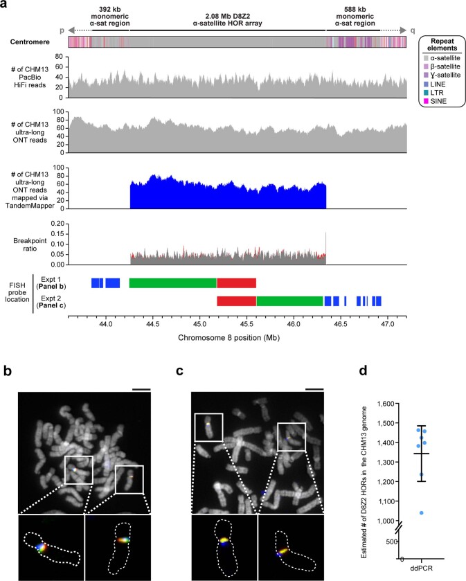
Extended Data Fig. 6. Validation of the CHM13 chromosome 8 centromeric region.
a, Coverage of CHM13 ONT and PacBio HiFi data along the CHM13 chromosome 8 centromeric region (top two panels) is largely uniform, indicating a lack of large structural errors. Analysis with TandemMapper and TandemQUAST52, which are tools that assess repeat structure via mapped reads (third panel) and misassembly breakpoints (fourth panel; red), indicates that the chromosome 8 D8Z2 α-satellite HOR array lacks large-scale assembly errors. Five different FISH probes targeting regions in the chromosome 8 centromeric region (bottom) are used to confirm the organization of the α-satellite DNA (b, c). b, c, Representative images of metaphase chromosome spreads hybridized with FISH probes targeting regions within the chromosome 8 centromere (a). Insets show both chromosome 8s with the predicted organization of the centromeric region. d, Droplet digital PCR of the chromosome 8 D8Z2 α-satellite array indicates that there are 1,344 ± 142 D8Z2 HORs present on chromosome 8, consistent with the predictions from an in silico restriction digest and StringDecomposer42 analysis (Methods). Mean ± s.d. is shown. Scale bar, 5 μm. Insets, 2.5× magnification.

