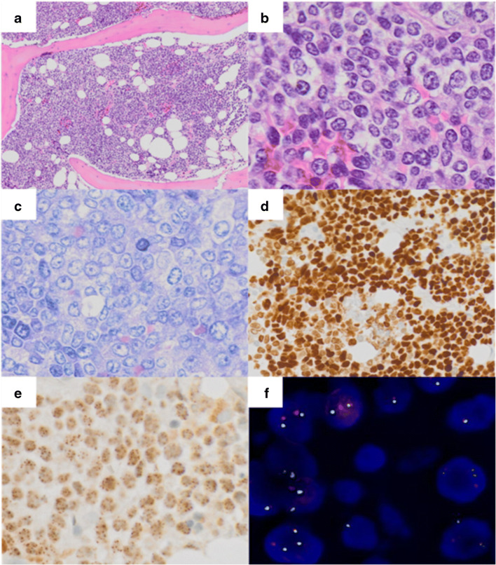
Fig. 1.
A–E Sheet-like proliferation of monomorphic cells infiltrating the bone marrow (a, × 10, Hematoxylin and eosin (HE)). Neoplastic cells feature a blastoid morphology with moderate amounts of clear cytoplasm and round to oval nuclei with vesicular chromatin (b, × 40, HE and c, × 40, Giemsa), staining for p63 (d, × 20, immunoperoxidase) and NUT (e, × 40, immunoperoxidase). f Dual-color FISH reveals a split-apart of the translocated NUTM1 gene

