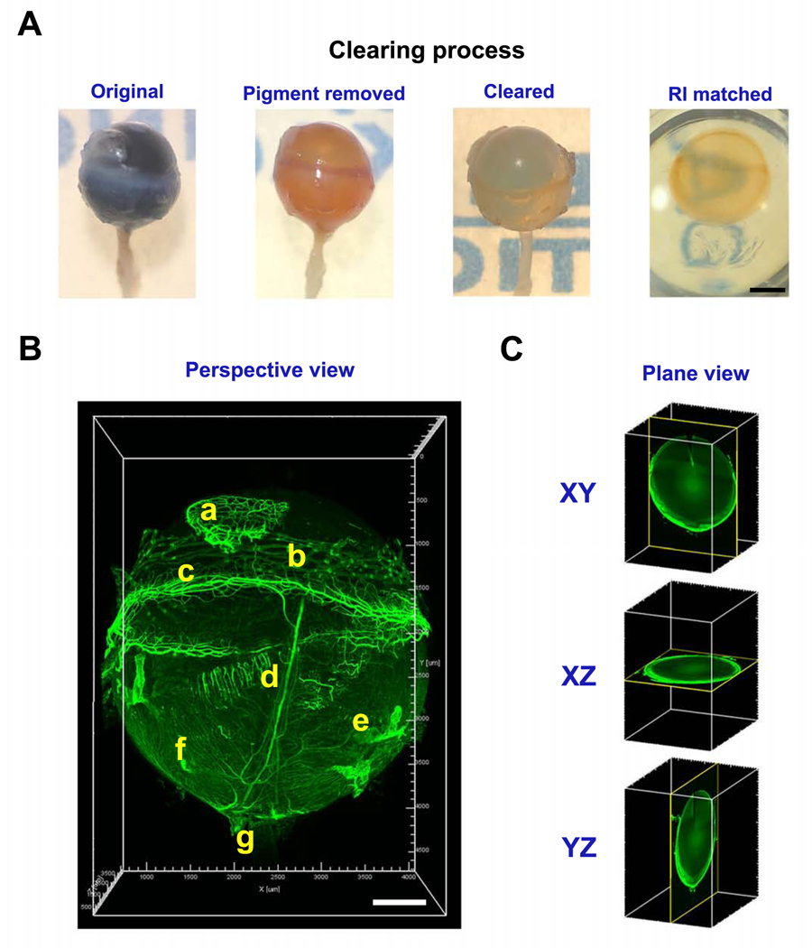
Figure 1. Representative images showing major clearing steps of a pigmented eyeball.
A. Stepwise images of a C57BL/6 mouse eyeball during tissue clearing process. RI: refractive index. B. Left panel, a 15° perspective view (all images unless otherwise indicated) of a cleared intact eyeball showing vasculature structures labeled by CD31 staining. a, nictitating membrane; b, conjunctiva; c, limbus; d, ciliary artery; e, vorticose vein; f, choroidal blood vessels; g, optic nerve. C. 2D views at different axial planes. Scale bar: 1 mm (A). 500 μm (B).

