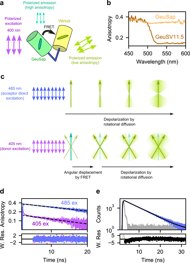
Fig. 3. GeuSV11.5 anisotropy.
a Cartoon representation of GeuSV11.5 under polarized excitation. b Steady-state anisotropy spectra of GeuSap and GeuSV11.5. c Top: schematic drawing of direct acceptor excitation of GeuSV11.5 by linearly polarized light. The yellow-green arrow represents the emission dipole moment of the acceptor. After absorption of polarized light, the polarization of the emitted light is gradually lost as a result of rotational diffusion. Bottom: donor excitation of GeuSV11.5 by linearly polarized light. FRET is accompanied by angular displacement from donor (green arrow) to acceptor (yellow-green arrows) dipole moments. Anisotropy of the sensitized acceptor reflects initially the angular displacement, followed by gradual depolarization due to rotational diffusion. d Time-resolved anisotropy of GeuSV11.5 under donor (405 nm; purple) or direct acceptor (485 nm; blue) excitation. The decay after 5 ns was fitted to a mono-exponential model, and yielded θ = 27 ns (sensitized acceptor) and θ = 34 ns (acceptor excited directly). e Fluorescence decay of GeuSV11.5 under 485 nm excitation (blue). The decay was fitted to a mono-exponential model with reconvolution (black) and yielded τ = 3.1 ns with χ2 = 1.29. W. Res.: weighted residuals. Source data are provided as a Source Data file.

