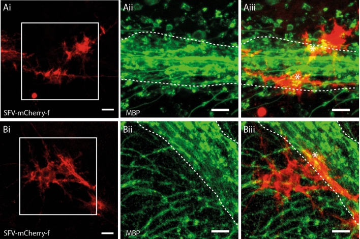
Figure 2.
SFV transduced astrocytes are morphologically and antigenically distinct from OL. (Ai,Bi) mCherry-f labelled glia in cerebellar white matter exhibiting complex finely branching process fields typical of astroglia. (Aii,Bii) Enlarged view of the boxed areas shown in (Ai)/(Bi) showing the major white matter paths (dashed white lines) with MBP+ OL processes (green) branching into the granule cell layer. (Aiii,Biii). Merged image showing mCherry-f fluorescence and MBP immunoreactivity from the same enlarged views shown in (Aii)/(Bii). White dashed lines indicate major white matter paths. In contrast to OL (Fig. 1) SFV labelled astrocytes lack cellular processes aligned in parallel with the major white matter tracks, or linear segments of MBP/process localization. Astrocytes exhibit patches of MBP co-localization (*) suggesting contact with MBP+ profiles within cerebellar white matter. Scale bars in all panels 20 µm.

