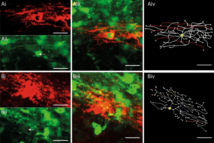
Figure 4.
Complement mediated injury destroys internodes on SFV labelled OL. (A) Reconstruction of an SFV labelled OL expressing the CNPase-GFP transgene in a complement control treated slice. (Ai) mCherryMBP fluorescence reveals an OL with a typical myelinating morphology. (Aii) CNPase-GFP signals imaged from the field shown in (Ai) (white arrow indicates position of the mCherry+ OL). (Aiii) Merged image of the field shown in (Ai-ii) confirms co-localisation of mCherry (red) and CNPase-GFP (green) signals. (Aiv) 3-D reconstruction and analysis reveals 4 internodes (red) on the OL shown in (Ai–iii). (B) Morphological analysis of an A774 labelled OL following complement injury. (Bi) mCherry-f labelled OL with degenerating processes. (Bii) CNPase-GFP signals imaged from the field shown in (Bi). White arrow indicates position of mCherry+ OL shown in (Bi). (Biii) Merged image reveals localization of mCherry (red) and CNPase-GFP (green) signals. (Biv) Reconstruction and analysis of the cell shown in (Bi–iii) reveals fragmented processes (coloured segments) and the absence of internodes. Yellow circles indicate position of cell body. Scale bars in (A) and (B) 20 µm.

