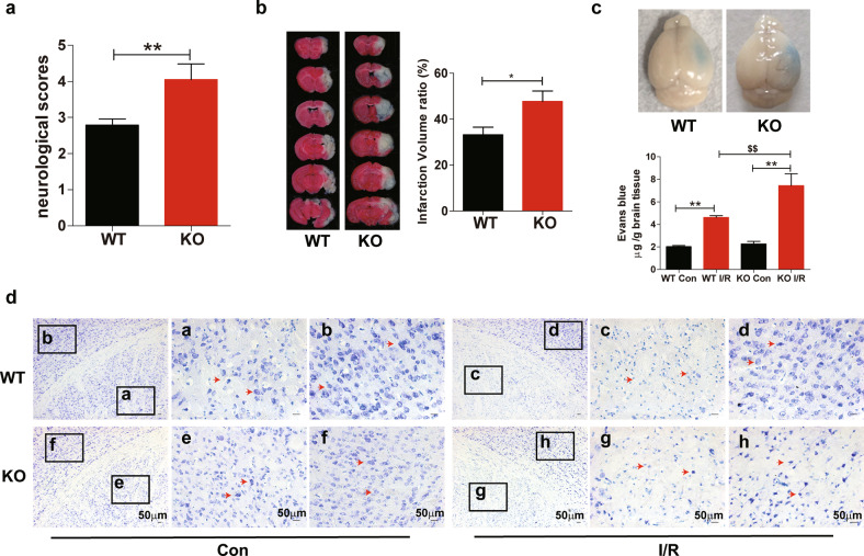
Fig. 2. Loss of FtMt exacerbates cerebral I/R-induced brain damage.
Wild-type and FtMt-knockout mice were subjected to MCAO for 90 min and subsequent reperfusion for 24 h. a After 24 h of reperfusion, the neurologic deficit scores of wild-type and Ftmt-knockout mice were compared (n = 15). b Infarct volumes were compared between wild-type (n = 5) and Ftmt-knockout mice (n = 6) by TTC staining of coronal sections. c Representative images (upper panels) and quantification (lower panels) of Evans blue dye extravasation in wild-type or Ftmt-knockout mice at 24 h after I/R (n = 5). d Nissl staining was performed after 90 min of MCAO and 24 h of reperfusion, and the I/R sides and control (Con) sides of wild-type and Ftmt-knockout mice were compared. The red arrows denote the altered Nissl bodies and degenerated neurons (n = 3). WT, wild-type mice; KO, Ftmt-knockout mice. The results are presented as the mean ± SEM. */$P < 0.05, **/$$P < 0.01.

