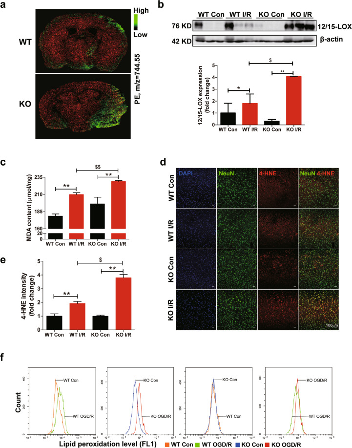
Fig. 4. Ablation of FtMt promotes lipid peroxidation in I/R brains.
a MSI heat maps for the distribution of PE (m/z = 744.55, green) in wild-type (WT) and Ftmt-knockout (KO) mouse brains after ischaemic stroke. b Western blot analysis of 12/15-LOX in WT and KO mice after MCAO (90 min) and subsequent reperfusion (24 h) (n = 6). c The mouse brain MDA content was measured. The data are expressed relative to the mean value in the WT control (Con) group (n = 5). d Representative immunofluorescence images of 4-HNE (red) and Neun (green). e Quantification of 4-HNE fluorescence intensity (n = 3). f Flow cytometry analysis of OGD/R-induced C11-BODIPY (581/591) oxidation in primary cultured WT and KO neurons. The data shown are representative of two independently performed experiments. The results are presented as the mean ± SEM. */$P < 0.05, **/$$P < 0.01.

