Figure 5.
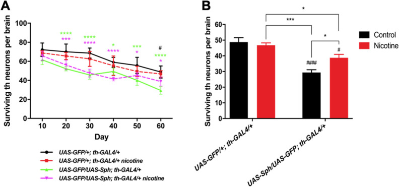
Nicotine promotes dopaminergic neuron survival. A) Dopaminergic neuron loss over time in control (UAS-GFP/+; th-GAL4/+) and experimental (UAS-GFP/UAS-Sph; th-GAL4/+) genotypes; untreated populations are represented by solid lines and treated populations by dotted lines. No significant differences were evidenced between the treated and untreated control genotype. Flies expressing Sph-1 in dopaminergic neurons have significantly fewer neurons than the control genotype throughout their life. However this difference becomes smaller between control and the Sph- expressing treated population as flies age. B) The number of surviving TH neurons in each experimental condition at the last time point studied (Day 60) evidences a reduction in th-positive neurons in the Sph-1 expressing flies compared with control genotype (#### p < 0.0001). Interestingly, this analysis also shows that nicotine treatment does not affect the number of th-positive neurons in the control genotype (p > 0.05), but it does so in the Sph-1 expressing flies (# p < 0.05 compared with the control genotype not treated with nicotine). The Sph-1 expressing flies treated with nicotine show a partial recovery in the number of th-positive neurons, since it is significantly increased compared with not-treated Sph-1 expressing flies (*p < 0.05), but does not reach the number of positive neurons recorded in the control genotype (*p < 0.05). In each case, data from controls were represented in black bars; data from animals treated with nicotine were represented in red bars. All statistical analysis were from two-way ANOVA followed by Tuckey’s post-hoc test, n = 5 fly brains. In all cases experimental animals were treated with 24 µM nicotine.

