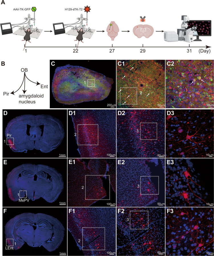
Fig. 6.
Our H129-dTK-T2-based tracing system enables visualization of detailed structures in postsynaptic neurons, aided by immunostaining enhancement of tdTomato signals. A Timeline for mapping the direct OB output pathways using H129-dTK-T2 along with AAV2/9-TK-GFP helper. Immunostaining against tdTomato is applied. B Schema of the simplified OB projection pathways. AAV2/9-TK-GFP, 1.0 × 10¹² vg/mL, 450 nL, H129-dTK-T2, 5.0 × 10⁸ pfu/mL, 400 nL, perfusion time: day 27. C Representative image of the virus injection site in the OB. The boxed areas are presented in the right panels at a higher magnification. The starter neurons labeled by both GFP and tdTomato are indicated by white arrows. D–F Representative labeling images of the regions innervated by the OB. To improve observation of the labeled post-synaptic neuron, the brain slice was immunostained with tdTomato antibody. The boxed areas are presented in the right panels at a higher magnification. Coronal brain slices were counterstained with DAPI, and tdTomato signals were amplified by immunostaining with the anti-DsRed polyclonal antibody and Alexa Fluor 594-conjugated goat anti-rabbit IgG (H+L). LEnt, lateral entorhinal cortex; MePV, medial amygdaloid.

