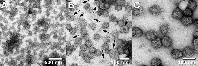
Figure 2.
Transmission electron micrographs of influenza virus A/Puerto Rico/8/34 H1N1 purified with steric exclusion chromatography (SXC) and pseudo-affinity chromatography with a sulfated cellulose membrane adsorber. Co-eluted exosome-like vesicles are visible in panels (A, B) and labeled with black arrows in panel (B). Panel (C) displays homogeneous (mostly intact) viral particles. Black arrows were added to the original published figure to highlight examples of extracellular vesicles (with lighter electron-density and collapsed cup shape) side by side with intact viral particles (non-collapsed round particles with higher electron density). All images are from the same sample at different magnifications. Pictures taken by Dietmar Riedel from the Max-Planck-Institute for Biophysical Chemistry in Göttingen, Germany. Reproduced with permission from Dr.-Ing. Pavel A Marichal-Gallardo, Max-Planck-Institute for Biophysical Chemistry in Göttingen, Germany (license CC BY-NC-SA 4.0).

