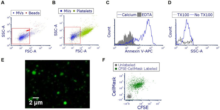
Figure 1.
Characterization of microvesicles (MVs) produced from pneumolysin (PLY)-treated alveolar epithelial cells. A549 cells were treated with PLY (100 ng/ml). After 4 h, conditioned media was collected and MVs were isolated and characterized. (A,B) Representative FACS analysis (overlay dot blots) of isolated vesicles (blue dots), and of (A) 1 μm beads (red dots) or (B) human platelets (green dots). (C) MVs were stained with annexin V-APC in the presence or absence of calcium and analyzed by FACS. (D) MVs were treated with Triton-X 100 and analyzed by FACS. (E) CFSE-labeled MVs (green) were visualized using confocal microscopy. (F) MVs were labeled with CFSE (FITC) and CellMask (APC) and analyzed by FACS. In the representative dot blot, MVs labeled with CFSE and CellMask are shown in green. Grey dots represent unstained vesicles (background staining).

