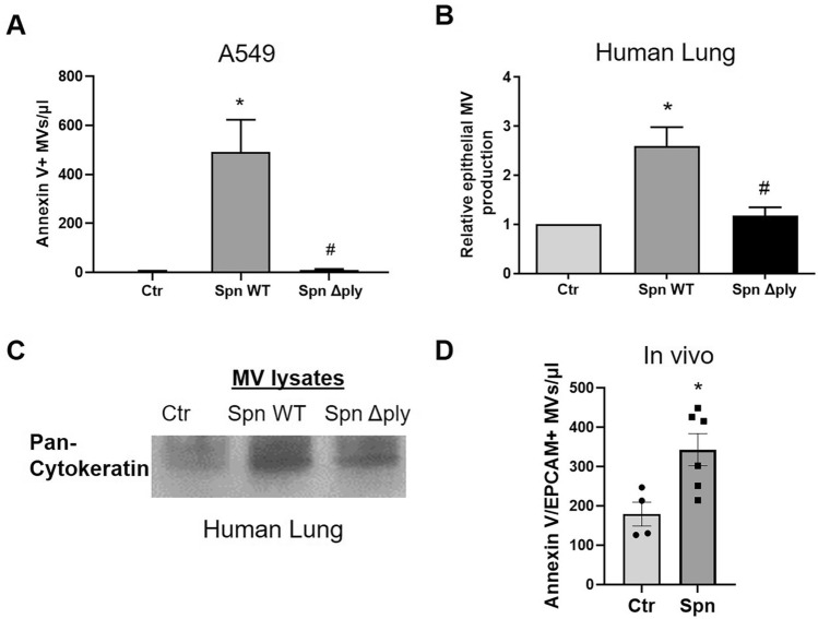
Figure 4.
Streptococcus pneumoniae (Spn) induces epithelial microvesicle (MV) production via pneumolysin. (A) A549 cells were treated with Spn WT, Spn Δply (MOI 50), or PBS (Ctr) for 4 h and MVs were isolated from conditioned media. Annexin V + MVs were quantified by FACS. N = 4 *p < 0.01 vs Ctr, #p < 0.01 vs Spn Δply (One-way Anova). (B,C) Human lung specimens (pieces from the same lung) were treated with Spn WT, Spn Δply (1 × 10⁶ CFU/ml), or PBS. (B) Graph depicts the relative amount of annexin V/EPCAM double positive MVs (normalized to Ctr). N = 6, *p < 0.05 vs Ctr, #p < 0.05 vs Spn ΔPly (One-way Anova). (C) MV lysates from human lung tissue supernatants probed for pan-cytokeratin (epithelial marker). Depicted is a representative blot (cropped image). (D) MVs of epithelial origin (annexin V/EPCAM double positive) were quantified in BAL of mice treated intranasally with Spn or PBS (Ctr). Each mouse is represented by a dot. N = 4–6, *p < 0.05 (unpaired t-test).

