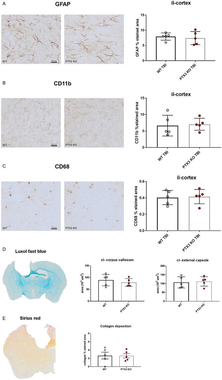
Figure 8.
PTX3 depletion did not modify brain inflammatory response at 5 weeks after TBI. Astrocytes (A, GFAP), brain myeloid cells (B, CD11b; C, CD68) and collagen deposition (E, by sirius red) were quantified in the quantified in the ipsilateral (il)-cortex within the first 350 μm from the edge of the contusion showing no difference between WT and PTX3 KO. Axonal loss in contralateral (cl)-corpus callosum and external capsule (D, by luxol fast blue) did not differ between the two strains. Data is presented as mean ± SEM, n = 5 (TBI WT/PTX3 KO). Unpaired t-test = ns.

