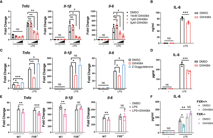
Figure 4.
FXR Agonist GW4064 Inhibits IL-6 Expression in Macrophages. (A, B) RAW 264.7 macrophages were incubated with or without different concentrations of FXR agonist GW4064 for 24h, and then stimulated with or without 100ng/ml LPS. (A) The mRNA levels of TNFα, IL-1β and IL-6 from harvested cells at 6h after the stimulation are shown. (B) IL-6 levels in the supernatant from 24-hr cultures are shown. (C, D) Bone marrow-derived macrophages were incubated in the presence of DMSO, 5μM GW4064 or 1μM FXR antagonist Z-Guggulsterone for 24h. Cells were then stimulated with or without 100ng/ml LPS. (C) The mRNA levels of TNFα, IL-1β and IL-6 from harvested cells at 6h after the stimulation are shown. (D) IL-6 levels in the supernatant from 24-hr cultures are shown. (E, F) Bone marrow-derived macrophages from FXR knockout (FXR−/−) mice and the littermates were incubated with or without 5μM GW4064 for 24h, and then stimulated with or without 100ng/ml LPS. (E) The mRNA levels of TNFα, IL-1β and IL-6 from harvested cells at 6h after the stimulation are shown. (F) IL-6 levels in the supernatant from 24-hr cultures are shown. Data are shown as mean ± SEM. Experiments were repeated 3 times and representative data are shown. *p < 0.05; **p < 0.01; ***p < 0.001; NS, no significant difference; # means compared to the relative unstimulated control group, two-way ANOVA, followed by Sidak’s multiple comparisons test.

