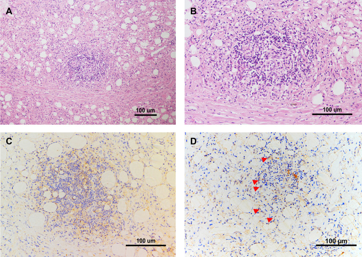
Figure 7.
IL-6 and FXR are Expressed in the Retroperitoneal Lesion of CP. (A, B) Representative images of HE staining in the biopsy of the retroperitoneal lesion from one CP patient. Scale bars: 100 μm. (C) Representative image of immunohistochemical staining of IL-6 in the biopsy of the retroperitoneal lesion. Scale bars: 100 μm. (D) Representative image of immunohistochemical staining of FXR in the biopsy of the retroperitoneal lesion. Scale bars: 100 μm. The red arrows indicate FXR positive cells especially within the nucleus.

