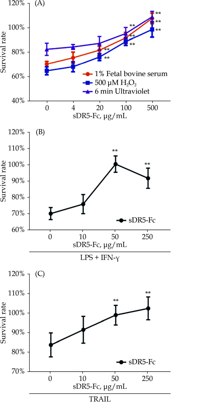
Figure 1.
sDR5-Fc recovered the proliferation of macrophage under extreme conditions in a dose-dependent manner.
All samples were exposed in different conditions for 24 h firstly and then treated with different concentrations of sDR5-Fc. (A): RAW264.7 cells were cultured in 1% fetal bovine serum (orange), 500 μM H2O2 (purple) or ultraviolet exposure (blue) separately; (B): RAW264.7 cells were induced with LPS and IFN-γ; and (C): RAW264.7 cells were supplemented with TRAIL. Data are expressed as mean ± SD from at least three representative experiments. **P < 0.01, compared with the group of sDR5-Fc with indicated concentration of 0 μg/mL. IFN-γ: interferon-gamma; LPS: lipopolysaccharide; sDR5-Fc: soluble death receptor 5-Fc; TRAIL: tumor necrosis factor-related apoptosis-inducing ligand.

