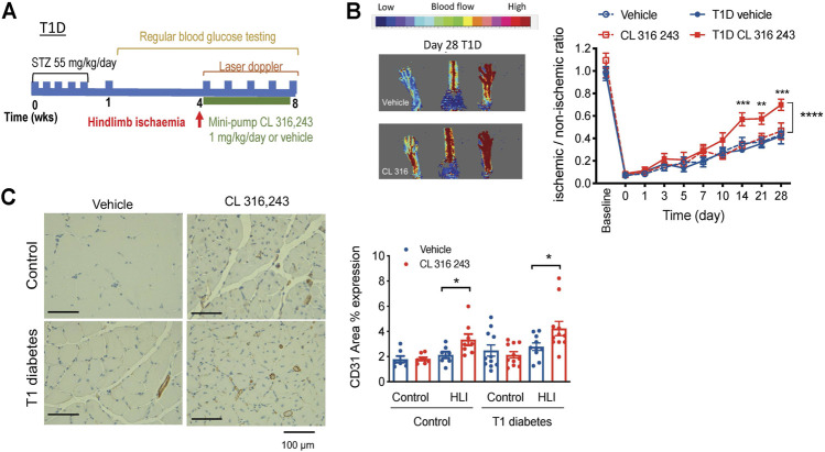
FIGURE 4.
Effect of β3AR stimulation in hind limb ischemia, as measured by laser doppler imaging, in type 1 diabetic mice. (A) Schematic diagram showing the study protocol. (B) Representative image in type 1 diabetic mouse at the end of the study (day 28) and (C) representative image of CD31 staining (left). Right, ratio of perfusion in ischemic to non-ischemic limbs in citrate-buffer control (n = 7–8) and type 1 diabetes (T1D, n = 10) mice treated with vehicle (saline) or CL 316,243 (1 mg/kg/day, s. c. 28 days). Mean ± SEM; *p < 0.05, **p < 0.01 ***p < 0.001 vs. vehicle by 2-way ANOVA with Bonferroni post-hoc analysis. (C) CD31 expression in hind limbs post-ischemia in control and diabetic mice treated with vehicle or CL 316,243. Representative images show CD31 stain in brown. Mean ± SEM; *p < 0.05, **p < 0.01 vs. vehicle by 1-way ANOVA with Bonferroni post-hoc analysis; n = 8–10.

