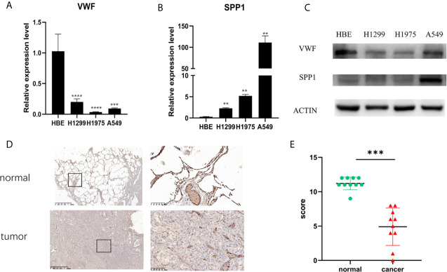
Figure 7.
Validation of mRNA and protein expression level. The mRNA expression level of VWF (A) and SPP1 (B) in HBE, H1299, H1975, and A549 cell lines. **p<0.01,***p<0.001,****p<0.0001. And protein expression of VWF and SPP1 validated using western blotting (C). Each experiment was carried out three times. A representative immunohistochemistry image of VWF in LUAD and adjacent non-tumor tissues. Scale bar = 100 µm (D). The scatter plot of immunohistochemistry score of LUAD and matched normal tissues. (E). ***p< 0.001.

