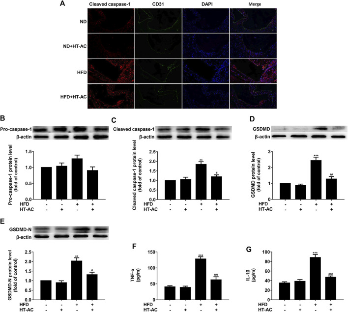
FIGURE 2.
HT-AC inhibited pyroptosis in the aortic intima of HFD-fed ApoE−/− mice (A) Frozen sections of aortic root were stained for activated caspase-1 (cleaved caspase-1) (stained in red). The nuclei were stained blue with DAPI. CD31 (stained in green) was used as an endothelial marker. Scale bar indicates 50 μm (B–E) The protein expressions of pro-caspase-1, cleaved caspase-1, GSDMD and GSDMD-N were determined by Western blotting (F,G) The concentrations of TNF-α and IL-1β in the serum were determined by ELISA. **p < 0.01, ***p < 0.001 vs. ND group; # p < 0.05, ## p < 0.01, ### p < 0.001 vs. HFD group. Results are expressed as mean ± SEM (n = 10).

