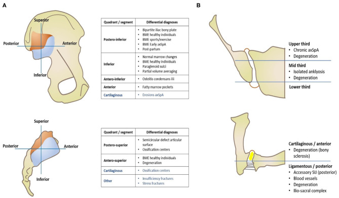
Figure 1.
Imaging of the sacroiliac joint—Topographic distribution of main anatomical variants and pathological conditions that mimic axSpA, separated by quadrants of each articular surface (A) and orthogonal planes (B), namely coronal oblique (right upper image) and axial oblique (right lower image).

