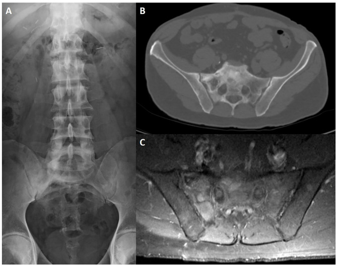
Figure 12.
Lumbosacral radiography (A), CT axial slice (B) and post-contrast fat-saturated T1WI (C) of a 65-year-old male patient with Paget disease of the sacrum and right iliac bone. Typical findings include an expanded bone with coarsened trabecular pattern and sclerotic changes that are more evident on conventional radiography. There is increased uptake after intravenous contrast injection (C).

