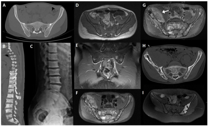
Figure 13.
CT axial slice (A) of an iliac bone enostosis mimicking peri-articular sclerosis; CT sagittal reconstruction (B) of the dorsal and lumbar spine in a patient with diffuse osteoblastic metastasis due to prostate cancer; lateral lumbar radiograph of the same patient (C); T1WI axial slice (D); and post-contrast fat-saturated T1WI coronal slice (E) of a patient with leukemic infiltration of the sacrum and iliac bones, showing diffuse bone marrow T1 hypointensity due to tumoral infiltration and multifocal patchy uptake, respectively; fat-saturated PD (F), post-contrast fat-saturated T1WI (G) MRI and CT axial slice (H) of a 18-year-old male patient with Ewing sarcoma; fat-saturated PD (I) MRI sequence of an aneurysmatic bone cyst of the left iliac bone.

