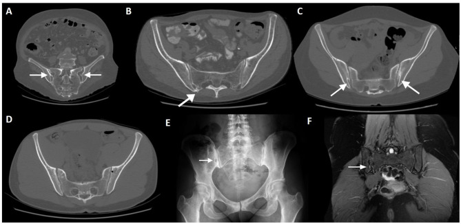
Figure 3.
Normal variants and incidental findings of the sacroiliac joint (arrows). CT reconstructions with oblique orientation depict bilateral iliosacral complexes (A), the most common sacroiliac joint variant; right accessory sacroiliac joint (B); bilateral bipartite iliac bony plate (C); left iliac bone pneumatocyst (D). A patient with an incidental finding on the right sacroiliac joint seen on pelvic radiography performed MRI, which revealed an iliac bone cleft filled with fluid (E,F). Note the sclerosis of the symphysis pubis (E), compatible with osteitis pubis.

