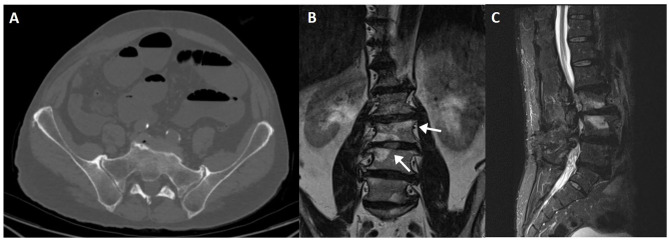
Figure 5.
CT axial slice showing degenerative changes of the sacroiliac joint (A), with marginal osteophytes and bone sclerosis. Modic endplate changes at the weight bearing surfaces of the distal lumbar spine (arrows), seen on coronal T1 (B) and sagittal fluid-sensitive (C) sequences. Bone marrow signal changes are high on T1WI and fat-saturated T2WI, compatible with Modic type 2.

