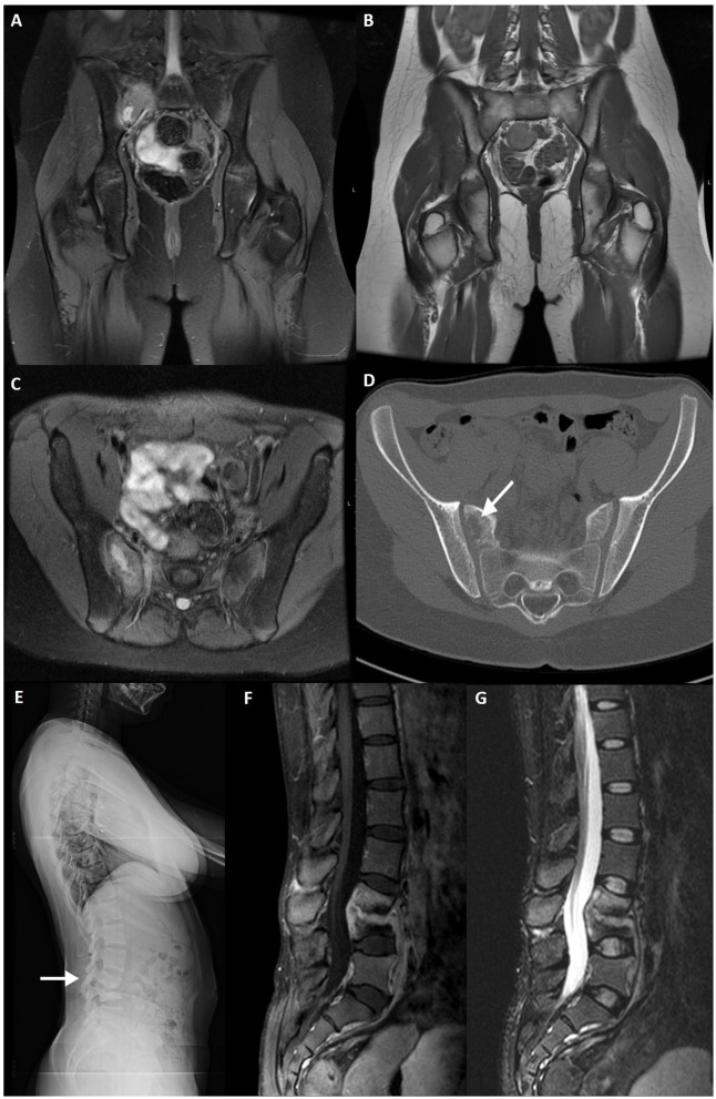
Figure 9.
Fat-saturated PD (A) and T1WI (B) coronal slices, fat-saturated PD (C) axial and CT axial slices (D) of a 12-year-old female patient with proven Streptococcus spp. osteomyelitis of the right sacrum (arrow). A lytic lesion is seen adjacent to the right sacroiliac joint. Lateral lumbar radiography (E), post-contrast fat-saturated T1WI (F) and TIRM (G) sagittal slices of a 26-year-old female patient with confirmed tuberculous spondylodiscitis of L3–L4 segment (arrow).

