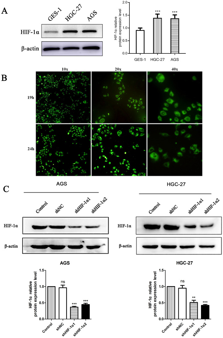
Figure 1.
(A) Western blot shows the HIF-1α expression level in gastric cancer cells and normal gastric epithelial cell (*P<0.001 compared with GES-1 group). (B) Fluorescence microscopy observed that DS entered the cytoplasm of human gastric cancer cells (HGC-27) 19 h and 24 h after DS intervention. (C) Western blot showed that the HIF-1α was successfully knockdown in human gastric cancer cells (ns, **P<0.01, ***P<0.001 compared with Control group).

