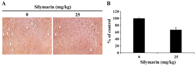
Figure 7.
Effects of silymarin on p-ERK1/2 expression in MCF-7 tumor tissues. (A) BALB/c nude mice were maintained under a 12-h light/dark cycle, and housed at controlled temperature (23±3°C) and humidity (40±10%) conditions. MCF-7 cells were injected subcutaneously into the right flank of donor nude mice, and silymarin was orally administrated 5 times per week at a dose of 25 mg/kg body weight for 3 weeks. Immunohistochemistry was conducted using antibodies against p-ERK (magnification, ×200). Arrows indicate p-ERK1/2-positive staining. (B) Graph presenting the quantification of p-ERK1/2 positive staining. *P<0.05 vs. untreated control (0 µg/ml silymarin). p-, phosphorylated.

