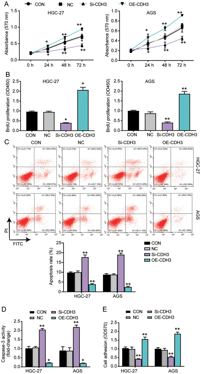
Figure 3.
Overexpression of CDH3 enhances the viability, proliferation and adhesion, and represses the apoptosis of gastric cancer cells. (A) Cell viability was detected in AGS and HGC-27 cells transfected with OE-CDH3 or Si-CDH3 using the MTT assay. (B) Cell proliferation was ascertained in HGC-27 and AGS cells transfected with Si-CDH3 or OE-CDH3 by BrdU ELISA assay. (C) Apoptosis was determined in AGS and HGC-27 cells transfected with Si-CDH3 or OE-CDH3 using the FITC apoptosis detection kit. (D) Apoptosis was established in HGC-27 and AGS cells transfected with Si-CDH3 or OE-CDH3 by caspase activity assay kit. (E) Cell adhesion was detected in HGC-27 and AGS cells transfected with Si-CDH3 or OE-CDH3 using a cell adhesion assay kit. Data are presented as the mean ± SD (n=3), and at least three independent tests were performed for each experiment. *P<0.05 and **P<0.001 vs. NC. CDH3, cadherin 3; Si-CDH3, small interfering RNA-CDH3; OE-CDH3, overexpression-CDH3; CON, blank control; NC, negative control of Si-CDH3 co-transfected with negative control of OE-CDH3; OD, optical density.

