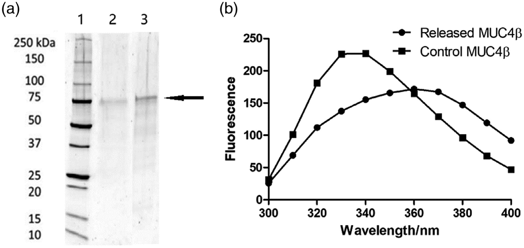
FIGURE 2.

Analysis of MUC4β structural stability. (a) SDS-PAGE of MUC4β released from nanoparticles. Lanes represent (1) molecular weight standard ladder; (2) released MUC4β; and (3) control MUC4β with 10 μg in each lane. The arrow indicates the location of the main MUC4β protein band. (b) Fluorescence spectroscopy of MUC4β released from nanoparticles. SDS-PAGE, sodium dodecyl sulfate polyacrylamide gel electrophoresis
