Fig. 1—

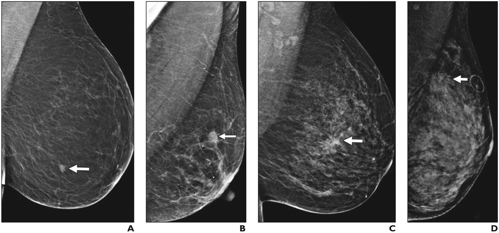
Left mediolateral oblique digital mammography (DM) images of cancer in breasts representing each of four BI-RADS breast density categories.
A, 61-year-old woman with pathogenic BRCA1 mutation. Screening DM image shows fatty breast and spiculated mass (arrow) that proved to be a 0.9-cm grade 3 invasive ductal carcinoma (IDC) that was estrogen receptor (ER) negative, progesterone receptor (PR) negative, and human epidermal growth factor receptor 2 (HER2 [also known as ERBB2]) negative with associated ductal carcinoma in situ (DCIS). Ki-67 proliferation index was high (90%). Three sentinel nodes were negative for metastasis.
B, 65-year-old woman with scattered fibroglandular breast tissue density. Screening DM image shows spiculated mass (arrow) that proved to be 1.1-cm grade 2 IDC with extensive DCIS that was ER positive, PR positive, and HER2 negative. Ki-67 proliferation index was high (30%), and metastatic sentinel node showed focal extracapsular extension.
C, 40-year-old woman with heterogeneously dense parenchyma, which may obscure small masses. Baseline screening DM image shows irregular mass (arrow) with distortion in central left breast. Core biopsy specimen showed 1.2-cm IDC that was ER positive, PR positive, and HER2 negative. Ki-67 proliferation index was moderate (15%). Patient went elsewhere for treatment.
D, 43-year-old woman with extremely dense parenchyma, which lowers sensitivity of mammography. Baseline screening DM image shows spiculated mass (arrow) with associated distortion in upper posterior left breast. This proved to be T2 (> 2 cm) grade 2 IDC with associated DCIS that was ER positive, PR positive, and HER2 negative. Ki-67 proliferation index was low (10%), and sentinel node biopsy was negative for metastasis.
