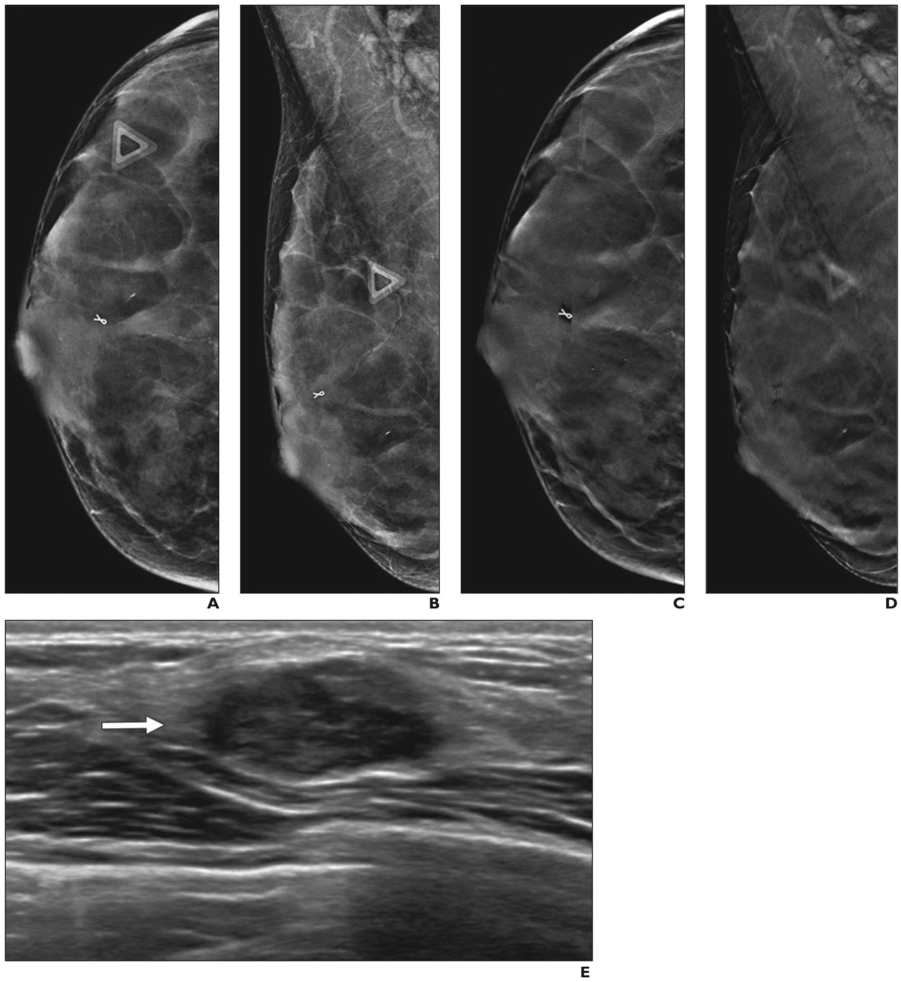
Fig. 3—

51-year-old woman with right breast pain who had cancer that was masked by extremely dense breast tissue on tomosynthesis.
A and B, Two-dimensional craniocaudal (A) and mediolateral oblique (B) digital mammograms show extremely dense parenchyma with ribbon clip from prior benign biopsy and no abnormality at site of pain (triangles).
C and D, Craniocaudal (C) and mediolateral oblique (D) digital breast tomosynthesis images show no abnormality.
E, Directed ultrasound image of area of focal pain shows 1.6-cm irregular, hypoechoic mass (arrow). Ultrasound-guided core needle biopsy revealed grade 3 invasive ductal carcinoma that was triple receptor negative. Excision after neoadjuvant chemotherapy showed no residual tumor. Sentinel node was negative for metastasis.
