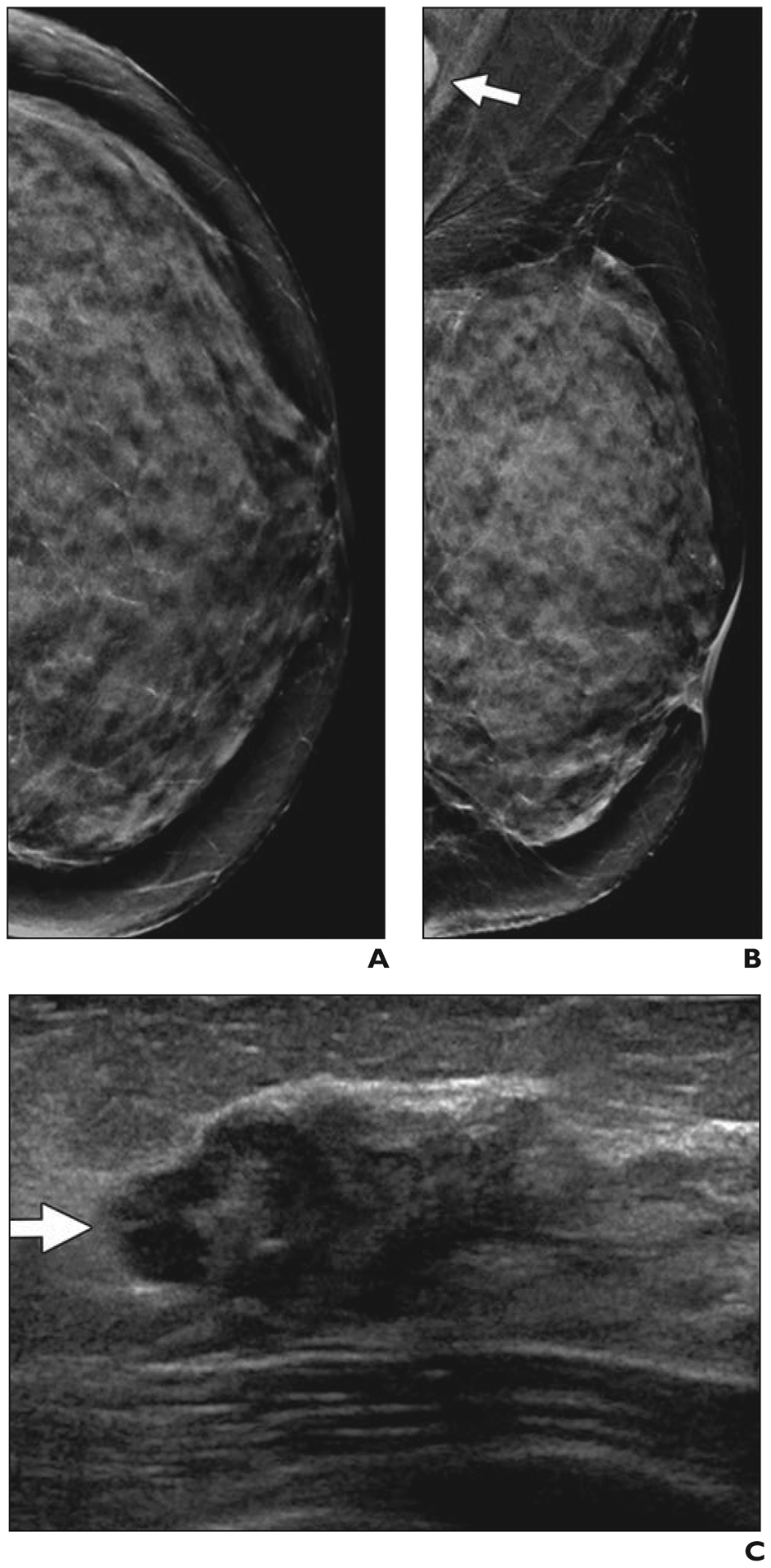
Fig. 4—

58-year-old woman with extremely dense breasts who had cancer seen on screening ultrasound (US) only.
A and B, Craniocaudal (A) and mediolateral oblique (B) synthetic 2D mammograms show normal extremely dense parenchyma, which was also interpreted as negative on digital breast tomosynthesis. In retrospect, portion of dense node is seen in left axilla (arrow, B). Handheld screening US performed by technologist was conducted bilaterally with standard documentation.
C, Radial US image of left breast shows irregular, hypoechoic 1.9-cm mass (arrow) located in 12-o’clock position 6 cm from nipple. US-guided core biopsy revealed grade 2 invasive ductal carcinoma that was estrogen receptor positive, progesterone receptor negative, and human epidermal growth factor receptor 2 (HER2 [also known as ERBB2]) amplified by fluorescence in situ hybridization. Ki-67 proliferation index was high (40%). US-guided core biopsy of left axillary node confirmed metastatic disease. Patient received primary chemotherapy and had no residual invasive carcinoma and few foci of ductal carcinoma in situ at lumpectomy. Targeted axillary dissection (with seed-localized excision of known metastatic node) showed one metastatic node with treatment effect and three normal nodes.
