Fig. 7—

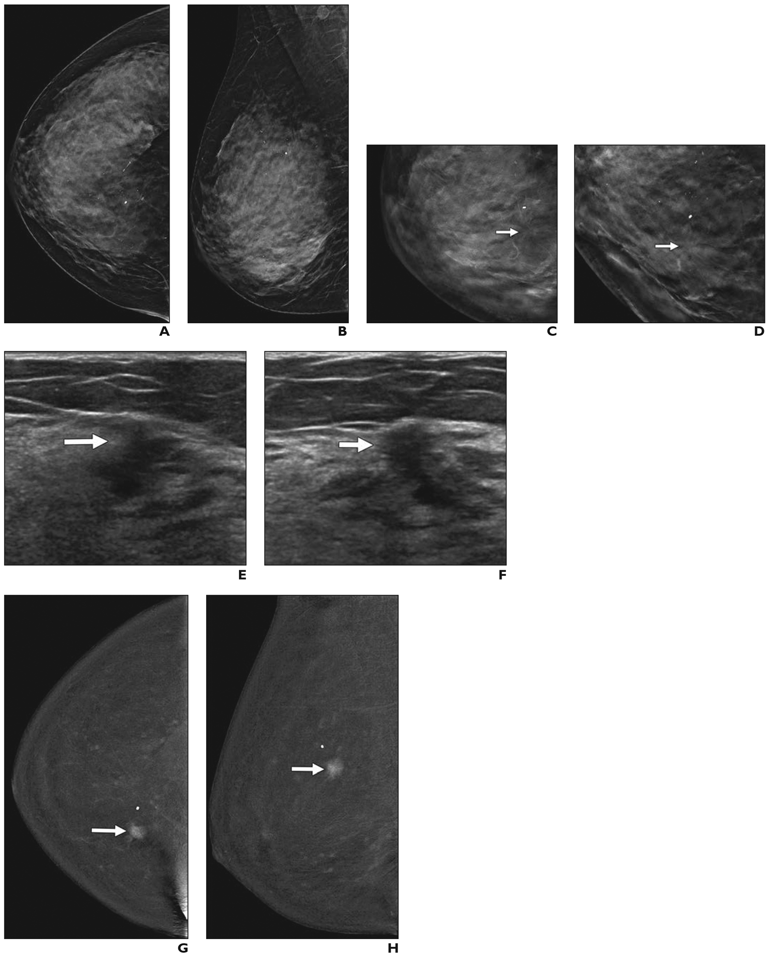
57-year-old woman with dense breasts who had invasive ductal cancer shown on contrast-enhanced mammography.
A and B, Screening craniocaudal (A) and mediolateral oblique (B) 2D synthetic mammography images show heterogeneously dense parenchyma and few calcifications.
C and D, Enlarged views of craniocaudal tomosynthesis image of inner right breast (C) and angled spot compression craniocaudal tomosynthesis image (D) show subtle distortion (arrows).
E and F, Targeted radial (E) and antiradial (F) ultrasound images of right breast show irregular, hypoechoic 0.9-cm mass (arrows) in 1-o’clock position located 6 cm from nipple, although findings of screening handheld ultrasound examination were negative. Before biopsy, patient underwent research contrast-enhanced mammography performed 2.5 minutes after IV injection of 125 mL of iopamidol (370 mg I/mL; Isovue 370, Bracco).
G and H, Craniocaudal (G) and mediolateral oblique (H) subtracted contrast-enhanced mammography images show strongly enhancing 1.2-cm irregular mass (arrows) and mild background parenchymal enhancement. Ultrasound-guided core biopsy revealed grade 2 invasive ductal carcinoma with ductal carcinoma in situ that was estrogen receptor positive, progesterone receptor positive, and human epidermal growth factor receptor 2 (HER2 [also known as ERBB2]) negative. Ki-67 proliferation index was low (10%). Invasive carcinoma measured 1.3 cm at lumpectomy, and four sentinel nodes were negative for metastasis.
