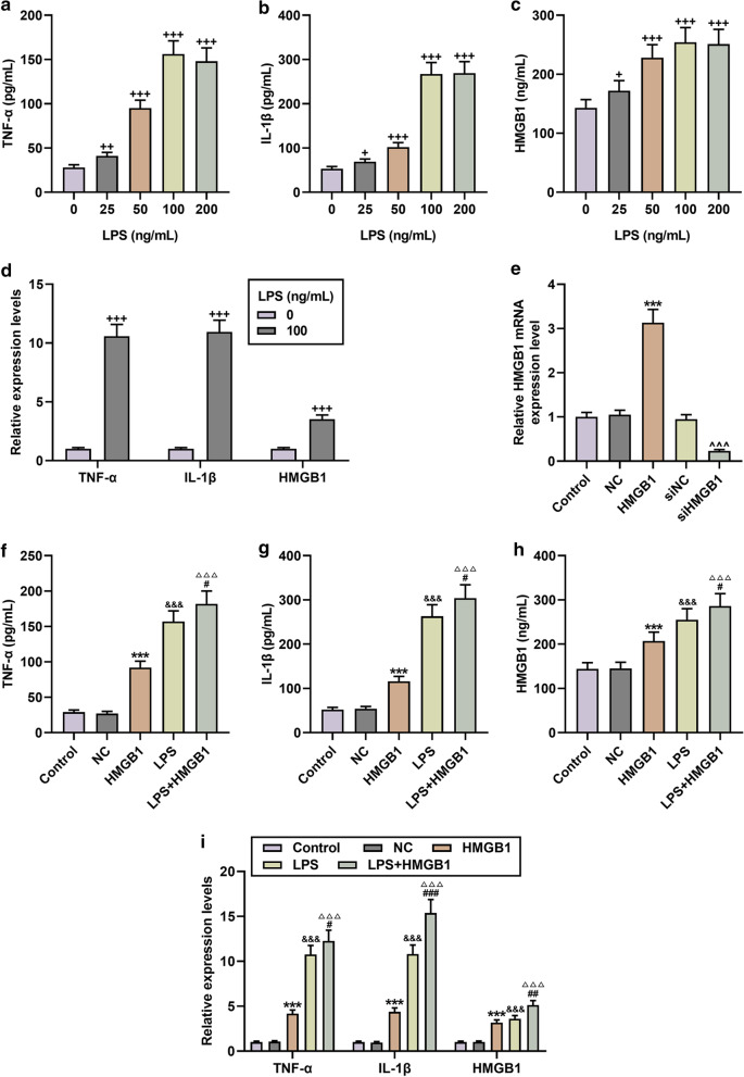
Fig. 1.
HMGB1 overexpression enhanced the levels of lipopolysaccharides (LPS)-stimulated cytokines in microglia. a–c After induction with different concentrations of LPS, the contents of TNF-α, IL-1β and HMGB1 in microglia were determined by ELISA. N = 3 for each column; +P < 0.05, ++P < 0.01, +++P < 0.001 vs 0; One-way ANOVA followed by Bonferroni's post-hoc test. d QRT-PCR was used to detect the expressions of TNF-α, IL-1β, HMGB1 in microglia cells stimulated or not stimulated by LPS. N = 3 for each column; +++P < 0.001 by t test. e The expressions of HMGB1 in the Control, Negative Control (NC), HMGB1 (HMGB1 over-expression vector), siNC, and siHMGB1 groups were determined by qRT-PCR. N = 3 for each column; ***P < 0.001, t test vs. NC; ^^^P < 0.001, t test vs siNC. (F-I) The results from ELISA and qRT-PCR showed that HMGB1 overexpression promoted the secretion and mRNA expressions of TNF-α, IL-1β and HMGB1, and enhanced the effect of LPS. N = 3 for each column; ***P < 0.001 vs. NC; &&&P < 0.001 vs. Control; △△△P < 0.001 vs. HMGB1; #P < 0.05, ##P < 0.01, ###P < 0.001 vs. LPS; One-way ANOVA followed by Bonferroni's post-hoc test. Each experiment was repeated three times independently. β-actin was used as a control. ELISA: enzyme linked immunosorbent assay; qRT-PCR: quantitative reverse transcription real time polymerase chain reaction

