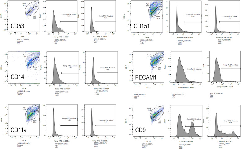
Fig. 5.
FACS scanning of EVs reveals a heterogeneous population of vesicles. UMSC-derived EVs were screened for microvesicle and exosome markers by FACS sorting. In the case of all vesicles, the EVs were separated into two populations, with the first population indicating higher side scatter than the second. Rightward peak shift indicates the presence of the stained markers. Population 1 showed a small increase in CD53+ (a), CD 151+ (b), CD14+ (c), and PECAM1+ vesicles (d). Population 2 showed a slight increase in CD151+ and CD14+ vesicles. Neither population showed an increase in CD11a+ vesicles (e). Finally, both populations had a strong presence in CD9+ vesicles (f)

