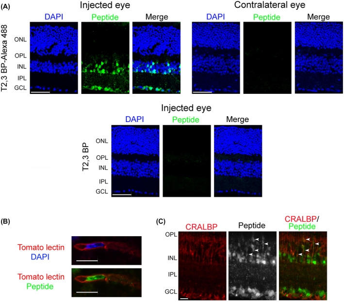
FIGURE 2.

ri CD40‐TRAF2,3 blocking peptide penetrates retinal cells. A, B6 mice received Alexa Fluor 488‐conjugated ri CD40‐TRAF2,3 blocking peptide or non‐fluorescent ri CD40‐TRAF2,3 blocking peptide (both 1 μg) via intravitreal injection of one eye. Injected and contralateral eyes were collected after 48 hours and frozen sections were examined. GCL = Ganglion cell layer; IPL = Inner plexiform layer; INL = Inner nuclear layer. OPL = Outer plexiform layer; ONL = Outer nuclear layer. Scale bar, 50 µm. B, C, Retinas from mice injected with Alexa Fluor 488‐conjugated ri CD40‐TRAF2,3 blocking peptide were stained with DyLight 594 tomato lectin (labels neural endothelial cells, B) or with anti‐CRALBP antibody (labels Müller cells, C). Green fluorescence was detected in cytoplasmic processes that co‐stain with CRALBP (arrowheads). Scale bar 10 µm. Original magnification 600× for panel B and 400× for panel C, Results are representative of three independent experiments
