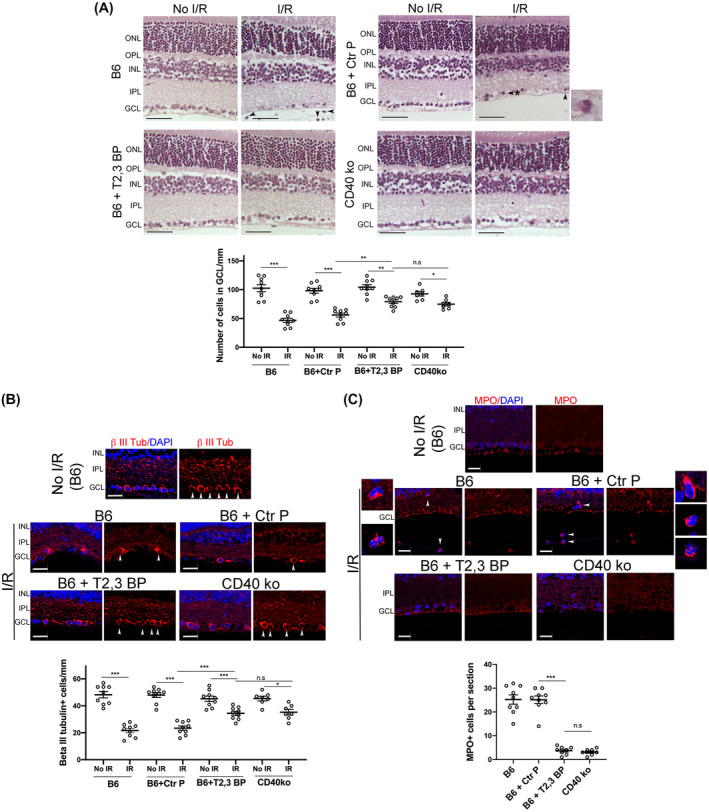
FIGURE 3.

ri CD40‐TRAF2,3 blocking peptide protects against cell loss in the GCL and infiltration by MPO+ leukocytes in retinas subjected to I/R. One eye from each B6 and Cd40−/− mouse was subjected to I/R. Contralateral non‐ischemic eye was used as control. Eyes subjected to I/R in B6 mice were treated intravitreously with or without ri control peptide (Ctr P), ri CD40‐TRAF2,3 blocking peptide (T2,3 BP; both 1 μg) 1 hour prior to an increase in IOP. Eyes were collected 2 days after I/R. A, Cell loss in the GCL is observed in ischemic eyes from B6 mice treated with ri control peptide or vehicle (original magnification 400×). H&E; Scale bar, 50 μm. Eyes from these mice also exhibited PMN infiltration in the inner retina and vitreous (arrowhead). Arrowhead plus asterix identifies a PMN magnified in the inset (original magnification 600×). The graph shows the numbers of cells in the GCL per mm. Horizontal bars represent mean ± SEM (9 mice per group). B, Sections were stained with anti‐β‐III tubulin antibody. Arrowheads identify β‐III tubulin+ cells. Original magnification 400×. Scale bar, 20 μm. The graph shows the numbers of β‐III tubulin+ cells in the GCL per mm. C, Sections were stained with anti‐MPO antibody. MPO+ cells (arrowheads) are magnified in the insets. Number of infiltrating MPO+ leukocytes in the inner retina and vitreous per section. No MPO+ cells were detected in the absence of I/R. GCL = Ganglion cell layer; IPL = Inner plexiform layer; INL = Inner nuclear layer. OPL = Outer plexiform layer; ONL = Outer nuclear layer. *P < .05; **P < .01; ***P < .001 by ANOVA
