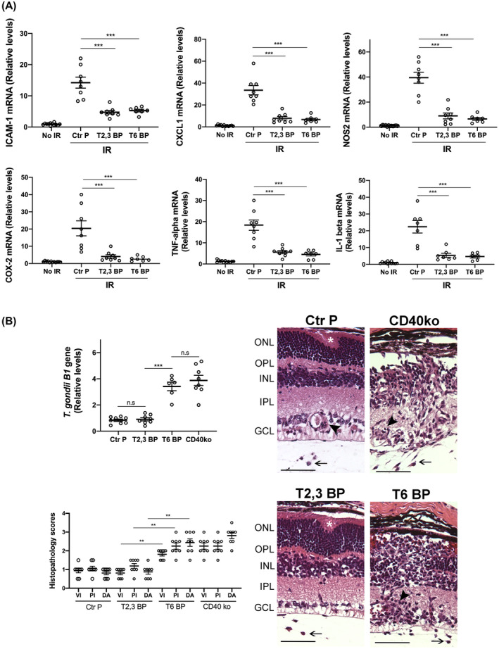
FIGURE 7.

Both the ri CD40‐TRAF2,3 and CD40‐TRAF6 blocking peptides impair retinal expression of inflammatory molecules induced by I/R but the ri CD40‐TRAF6 blocking peptide exacerbates infectious retinitis. A, Eyes subjected to I/R were treated intravitreously with or without ri control peptide (Ctr P), ri CD40‐TRAF2,3 blocking peptide (T2,3 BP) or ri CD40‐TRAF6 blocking peptide (T6 BP; 1 μg) 1 hour prior to an increase in IOP. Eyes were collected 2 days after I/R. mRNA levels of were assessed by quantitative real‐time PCR as above. B, B6 and Cd40−/− mice were infected with T. gondii tissue cysts. B6 mice received peptides intravitreally 4 days after infection. Eyes were collected 14 days post‐infection. Levels of T. gondii B1 gene expression were assessed by real‐time PCR. Eyes from infected B6 mice that received the CD40‐TRAF6 blocking peptide or infected Cd40−/− mice showed prominent disruption of retinal architecture (asterix), perivascular (arrowhead), and vitreal inflammation (arrow). H&E; X200. Bar, 50 μm. Histopathology scores for vitreal inflammation (VI), perivascular inflammation (PV), and disruption of retinal architecture (DA). Graphs represent mean ± SEM of 8 mice per group. **P < .01; ***P < .001 by ANOVA
