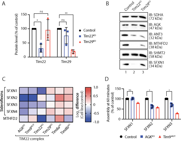
FIGURE 5:
Sideroflexins require the TIM22 complex for their biogenesis. (A) Mitochondrial lysates from triplicate sets of control, Tim22 knockdown (KD), and Tim29 KD HEK293 cells were analyzed by SDS–PAGE and Western blotting with antibodies specific for Tim22, Tim29, and SDHA (loading control). Levels of Tim22 and Tim29 were quantified and tabulated as mean ± SD (n = 3). One-sample t test: *, p < 0.05, **, p < 0.01. (B) Mitochondrial lysates from control, Tim22 KD, and Tim29 KD HEK293 cells were analyzed by SDS–PAGE and Western blotting. (C) Log2 fold-change values (as compared with respective controls) are depicted for selected proteins in the indicated cell lines. (D) [35S]-SFXN1, [35S]-SFXN2, and [35S]-SFXN3 were incubated with mitochondria isolated from control, AGKKO, and Tim9MUT HEK293 cells for 60 min before proteinase K (PK) treatment. Samples were solubilized in 1% digitonin containing buffer and analyzed by BN-PAGE and autoradiography. Assembled protein at 60 min in control, AGKKO, and Tim9MUT mitochondria was quantified. Graph depicts mean ± SD (n = 3 for AGKKO, n = 1 for Tim9MUT). One-sample t test: *, p < 0.05.

