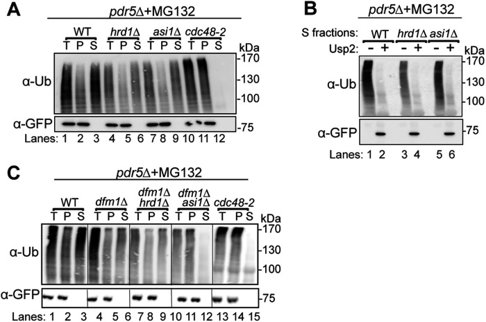
FIGURE 4:
Retrotranslocation of full-length Sec61-2-GFP. (A) In vivo retrotranslocation of Sec61-2-GFP through both Hrd1 and Asi channels. WT, hrd1Δ, asi1Δ, and cdc48-2 strains expressing Sec61-2-GFP were grown into log phase and treated with MG132 (25 µg/ml). Crude lysates were ultracentrifuged to separate Sec61-2-GFP that has been retrotranslocated into the soluble fraction (S) and Sec61-2-GFP that has not been retrotranslocated from membrane (P). Sec61-2-GFP was immunoprecipitated from both fractions and then analyzed by SDS–PAGE and immunoblotting with α-GFP and α-ubiquitin. One representative of three biological replicates is shown. (B) In vivo retrotranslocated Sec61-2-GFP is full length. WT, hrd1Δ, asi1Δ, and cdc48-2 strains expressing Sec61-2-GFP were grown into log phase and treated with MG132 (25 µg/ml). Crude lysates were ultracentrifuged to separate Sec61-2-GFP to collect retrotranslocated Sec61-2-GFP from soluble fractions. Solubilized Sec61-2-GFP was immunoprecipitated and then either treated with either buffer (–) or the catalytic core of the deubiquitinase Usp2 (+). Samples were analyzed by SDS–PAGE and immunoblotted with α-GFP and α-ubiquitin. One representative of three biological replicates is shown. (C) In vivo retrotranslocation of Sec61-2-GFP through Asi1 is Dfm1 independent. WT, dfm1Δ, dfm1Δhrd1Δ, dfm1Δasi1Δ, and cdc48-2 strains expressing Sec61-2-GFP were grown into log phase and treated with MG132 (25 µg/ml). Crude lysates were ultracentrifuged to separate Sec61-2-GFP that has been retrotranslocated into the soluble fraction (S) and Sec61-2-GFP that has not been retrotranslocated from membrane (P). Sec61-2-GFP was immunoprecipitated from both fractions and then analyzed by SDS–PAGE and immunoblotting with α-GFP and α-ubiquitin. One representative of three biological replicates is shown.

