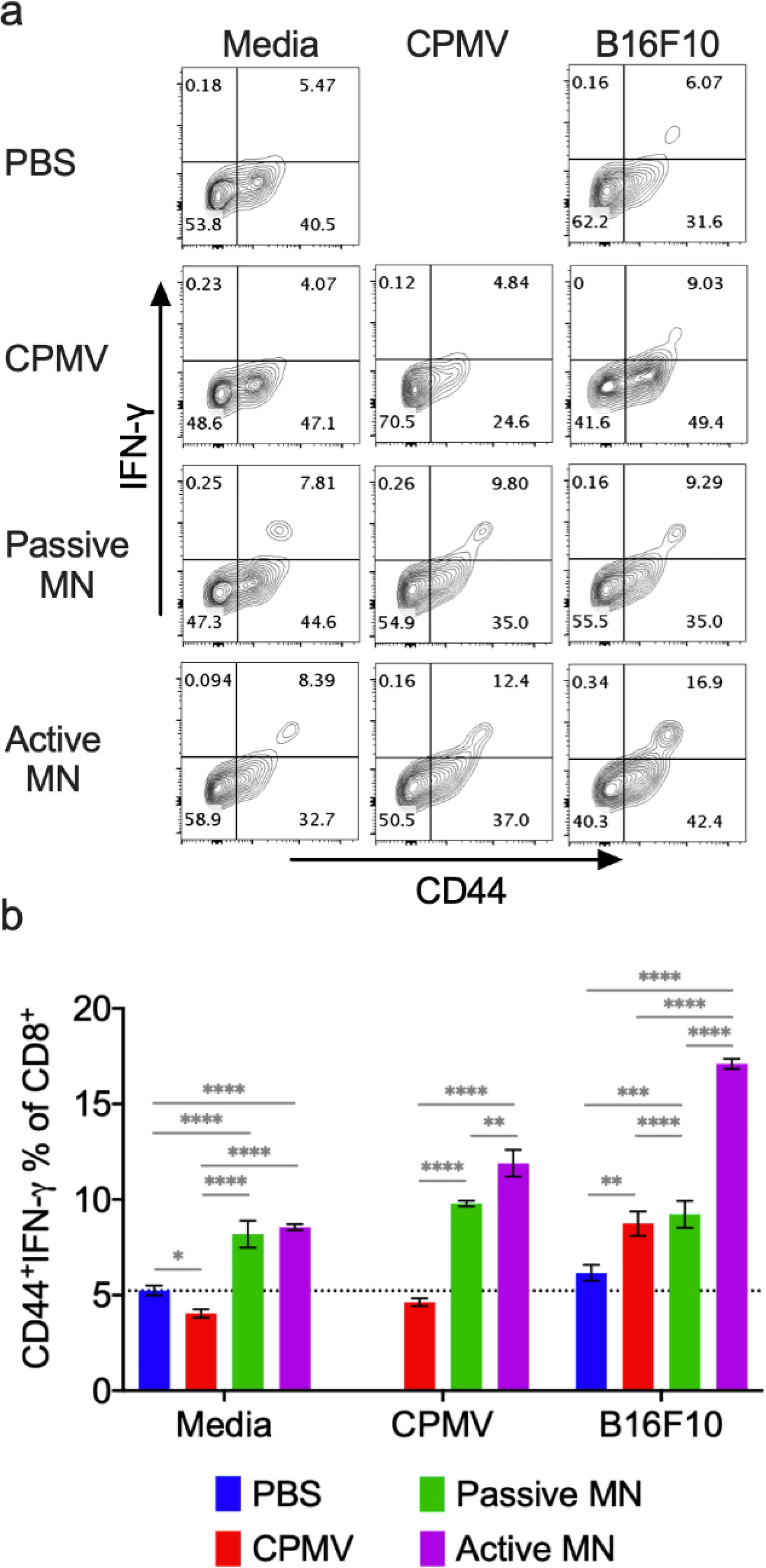
Figure 6. Systemic anti-tumor immune response following CPMV microneedle administration.

C57BL/6 mice bearing dermal B16F10 tumors (60 mm3) were treated with CPMV by intratumoral injection, passive MN, or active MN 10 days after B16F10 cell inoculation. 10 days following the treatment, spleens were harvested and co-cultured with media, 10 μg CPMV, or B16F10 tumor cell lysate for 48 h. Intracellular IFN-γ was measured in CD8+ T cells by flow cytometry. (a) Representative flow cytometry plots of CD44hiIFN-γ+CD8+ T cells in each re-stimulation group. (b) The percentage of CD44hiIFN-γ+CD8+ T cells after gating CD8+ T cells. Data are means ± SD (n=3). The dashed line indicates the background level activation. Splenocytes from PBS-treated animals were omitted from CPMV stimulation. Statistical significance was calculated using one-way ANOVA with Tukey’s multiple comparisons post-test: **P<0.05, **P<0.01, ***P<0.001, ****P<0.0001.
