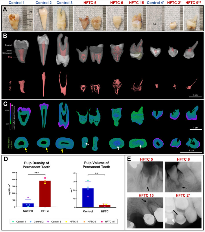
Fig 3.

Clinical images and three‐dimensional (3D) reconstruction of the teeth of patients with hyperphosphatemic familial tumoral calcinosis (HFTC) reveal abnormal root and pulp. (A) Images of selected control and HFTC teeth samples. HFTC teeth have shorter roots and more irregular root surfaces. (B) 3D reconstructions with segmentation (see Patients and Method section) of control and HFTC teeth are shown (white = enamel, gray = dentin/cementum, pink = pulp, yellow = composite buildup). Pulp (pink) from 3D reconstructions shown in (B) are highlighted. Although control teeth have single pulp chambers, HFTC teeth show reduced pulp chamber and canals with capillary‐like morphology. (C) Longitudinal and cross‐sectional heat maps illustrate differences in density distribution. Color bar shows increasing density (green to purple). In HFTC teeth, dentin and cementum layers are indistinguishable. Areas of high density are found towards the periphery of root in control (yellow arrows) versus towards the core in HFTC teeth (white arrows). The majority of pulp has been replaced in HFTC teeth. Density of primary HFTC teeth does not appear to be as severely affected. (D) The mean pulp density was sevenfold higher in permanent HFTC teeth than in controls (382 ± 39 vs 53 ± 58 mg HA/cm3, p = 0.0004). The mean pulp volume was sevenfold lower in permanent HFTC teeth than in control (3 ± 1 vs 22 ± 8 mm3, p = 0.0088). (E) Periapical radiographs of tooth #30 from patient 5, tooth #21 from patient 6, tooth #5 in patient 15, and tooth C in patient 2. Periapical radiolucency, pulp obliteration, and root bulging are observed. Black arrows indicate extracted/exfoliated teeth. *Primary teeth. †Patient 9 has a triplication within chromosome 13 likely affecting KLOTHO.
