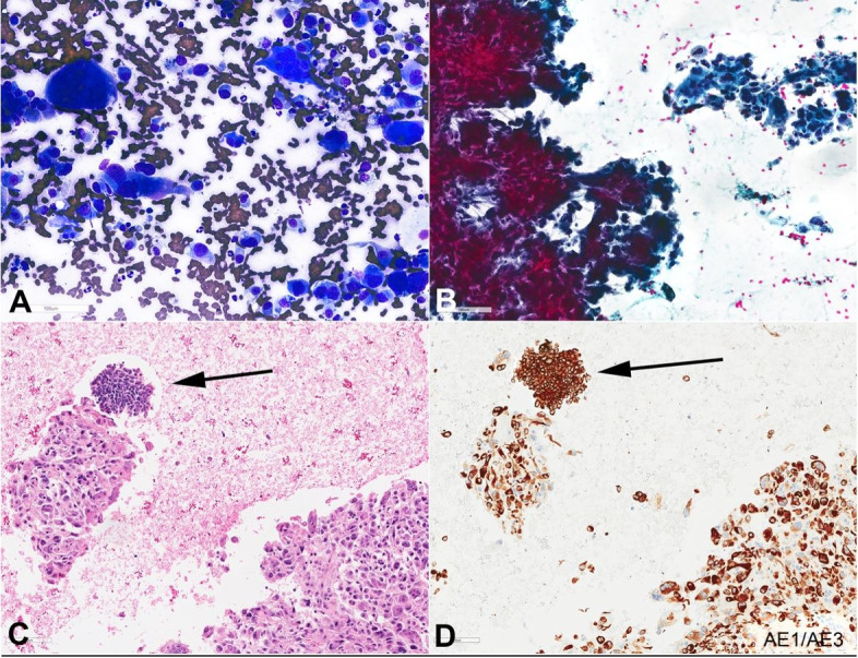
Figure 2. Photomicrographs of endoscopic ultrasound (EUS)-guided fine-needle aspiration biopsy (FNA) of the pancreas mass (400 x magnification); A – Diff-quick-stained smear showing hypercellular smear consists of pleomorphic tumor cells and osteoclast-like giant cells; B – Papanicolaou-stained smear showing hypercellular smear consists of cohesive spindle to pleomorphic tumor cells and osteoclast-like giant cells; C – H&E section of the cell block show pleomorphic and spindle cell neoplasm with scattered osteoclast giant cells (UCOGC component). Foci of smaller neoplastic round to spindle cells with scant finely granular cytoplasm and “salt and pepper” (finely stippled) chromatin are noted (neuroendocrine component; arrow); D – AE1/AE3 immunostain.

