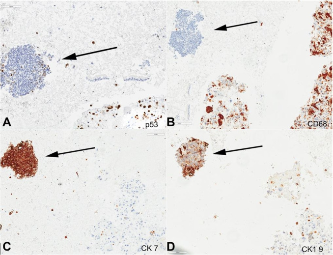
Figure 3. Photomicrographs of endoscopic ultrasound (EUS)-guided fine-needle aspiration biopsy (FNA) of the pancreas mass (400 x magnification), arrow indicates neuroendocrine component and the remaining cells are UCOGC component; A – P53 immunostain; B – CD68 immunostain; C – CK7 immunostain; D – CK19 immunostain.

