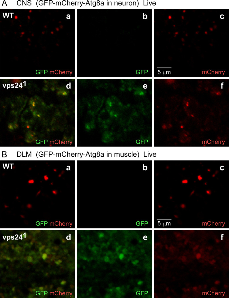
Fig 9. The GFP-mCherry-Atg8a fusion proteins accumulate in a non-acidic compartment in the vps24 mutant.
Confocal live-imaging of the GFP-mCherry-Atg8a fusion protein in CNS neurons (A) and DLM (B) from WT (a-c) or the vps24 mutant (d-f). Neuronal (A) or muscle (B) expression of the tandem-tagged fusion protein was achieved as described in Fig 2. In both neurons and muscle of the vps24 mutant, colocalized GFP and mCherry fluorescence was detected (Ad-f, Bd-f). This indicates that the pH-sensitive GFP tag is not quenched and thus the tandem-tagged fusion protein is in a non-acidic compartment. In WT neurons and muscle (Aa-c, Ba-c), the GFP fluorescence signal is diminished with respect to the mCherry fluorescence.

