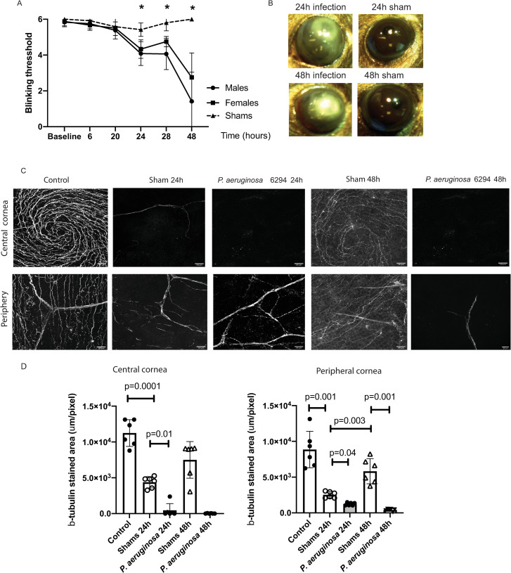
Fig 1. Bacterial infection induces neuropathy.
A. Corneal sensitivity was tested using Cochet-Bonnet esthesiometer. Infected male (N = 6) and female (N = 6) cohorts of C57BL6/N mice were compared to shams (N = 6). Changes in the blink reflexes were monitored over time (hours). The asterisks denote significant differences in blink reflexes (Two-way ANOVA, 24h, p = 0.0001; 28h, p = 0.0006; 48h, p = 0.0001). B. Eyes from C57BL6/N mice were either scratched (shams) (N = 6) or scratched and infected with 5x10⁵ CFU/eye P. aeruginosa 6294 (N = 6). Representative images of eye appearance at 24h and 48h post-infectious challenge are shown. The images were acquired using Motic SMZ140-143 stereomicroscope. C. Corneal tissues were harvested at 24h and 48h after challenge and corneal tissues were stained for β-tubulin. Z-stacks of corneal whole-mounts covering a depth of 40 μm were acquired with 1 μm step. The z- stacks were collapsed to 2D. The images are representative 2D projections. The top series of images are depicting central area of corneas while the bottom panels depict β-tubulin staining in peripheral corneas; scale bar, 20 μm. the appearance of six individual corneal whole mounts were compared per condition. D. Image quantification using Fiji. The 40 μm z-stacks were collapsed to 2D and areas of β-tubulin staining were calculated per slide. Data are presented with bar plots where each symbol represents the value of an individual animal. One-way ANOVA, overall p = 0.001. P values for the individual comparisons are shown on the plot. Asterisks indicate significant differences. Data demonstrate that infection changes corneal pain sensation and reduces densities of the neuronal network in central and peripheral corneas.

